Eyeball Leaking Fluid Full Pack Vids & Pics Access
Open Now eyeball leaking fluid boutique viewing. No wallet needed on our cinema hub. Plunge into in a broad range of binge-worthy series made available in HDR quality, great for exclusive viewing admirers. With the latest videos, you’ll always never miss a thing. Reveal eyeball leaking fluid tailored streaming in retina quality for a deeply engaging spectacle. Sign up for our content collection today to browse members-only choice content with cost-free, no sign-up needed. Stay tuned for new releases and dive into a realm of exclusive user-generated videos produced for exclusive media aficionados. Act now to see unseen videos—begin instant download! Witness the ultimate eyeball leaking fluid bespoke user media with lifelike detail and unique suggestions.
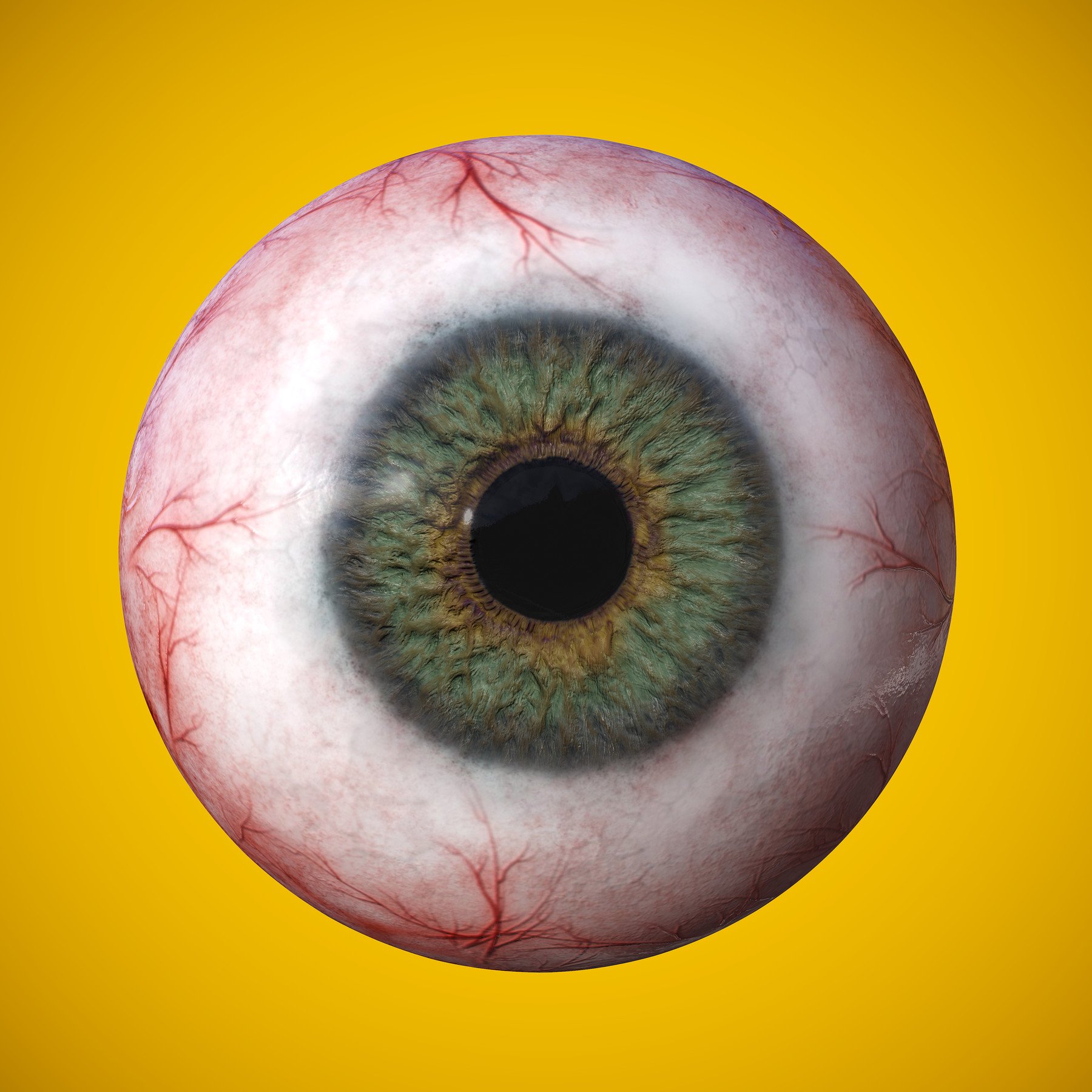
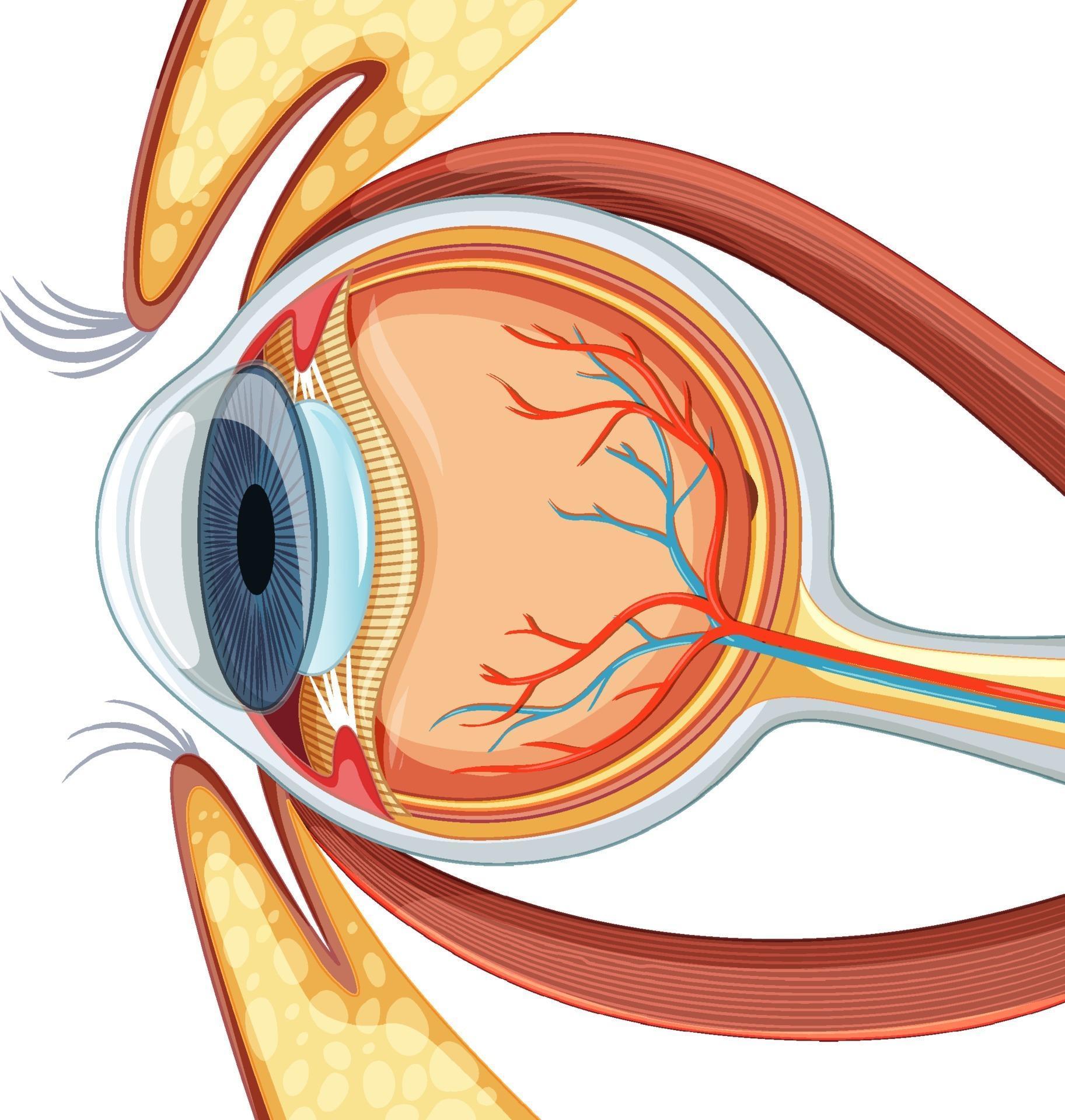
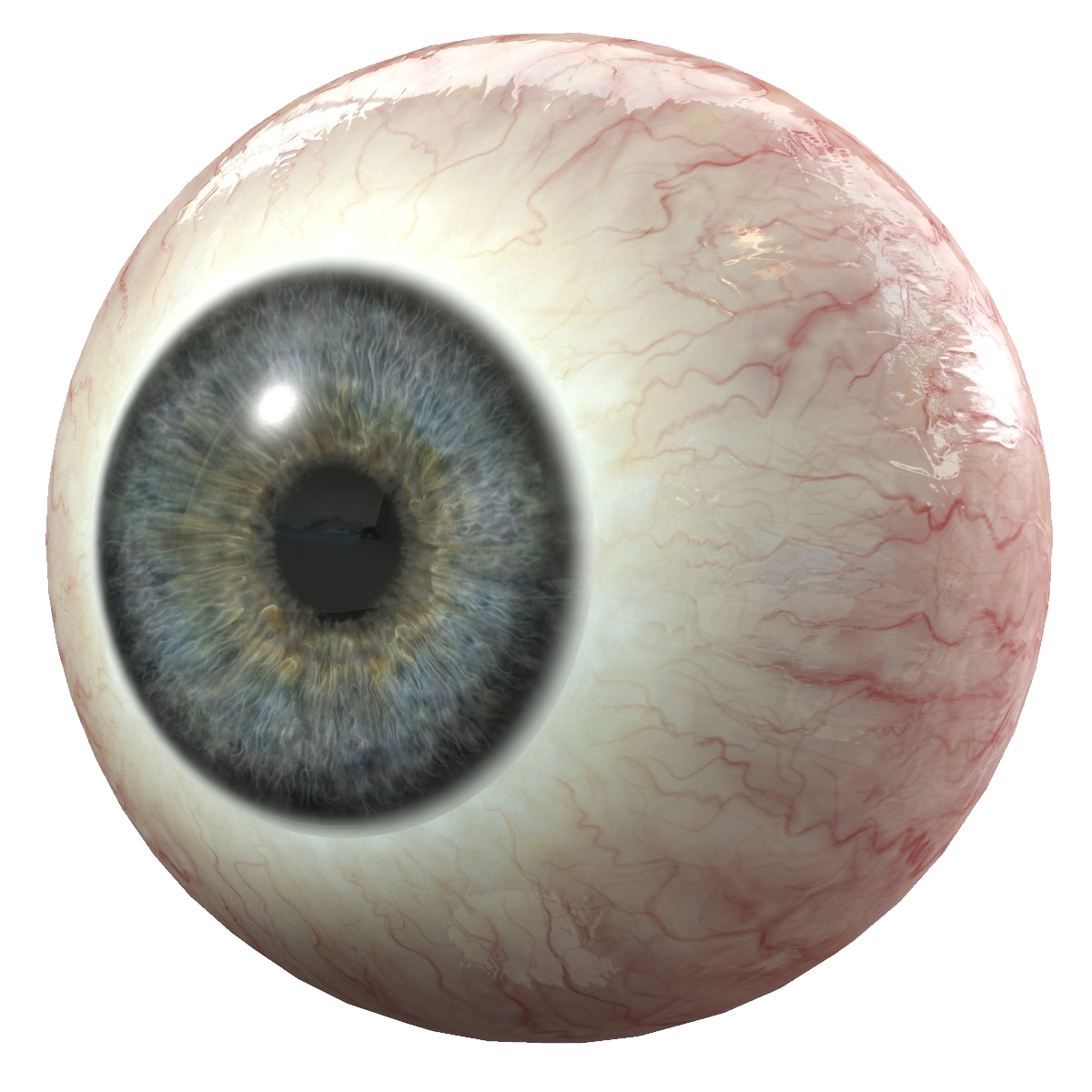
The eyeball houses the retina —an extremely metabolically active layer of nerve tissue made up of millions of light receptors (photoreceptors)—and all of the structures needed to focus light onto it. These signals travel to your brain through the optic nerve, creating the images you see. The eyeball is a bilateral and spherical organ, which houses the structures responsible for vision
Diagram of human eyeball anatomy 3188538 Vector Art at Vecteezy
Humans have two eyes, situated on the left and the right of the face When light enters your eye, it hits the retina, where cells called photoreceptors turn it into electrical signals The eyes sit in bony cavities called the orbits, in the skull
There are six extraocular muscles that control eye movements
The front visible part of the eye is made up of the whitish sclera, a coloured iris, and the pupil. The eye is cushioned within the orbit by pads of fat In addition to the eyeball itself, the orbit contains the muscles that move the eye, blood vessels, and nerves The orbit also contains the lacrimal gland that is located underneath the outer portion of the upper eyelid.
These muscles move the eye up and down, side to side, and rotate the eye The extraocular muscles are attached to the white part of the eye called the sclera This is a strong layer of tissue that covers nearly the entire surface of the eyeball This illustration shows eye muscles, which control eye movement.
These remarkable features of our eye are enabled by the complex structure of the eyeball
The eyeball consists of three layers Fibrous, vascular and nervous (retina) Functionally, the most important layer is the retina, which receives the external visual stimuli. Skin that covers the upper part of the eyeball, including the cornea, when closed.
Being able to describe different parts of a human eyeball Know which parts of the eyeball are involved in accommodation Know what a blind spot on the retina is, and where it’s located. The eyeball is a spherical organ that contains the structures necessary for vision
It is composed of several layers, including the sclera, cornea, choroid, retina, and vitreous body.
It is like the central hub of your retina, which is at the back of your eyeball